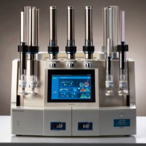
 Customization of flow cytometry panels is far from being a matter of preference or opulence, it stands as a requirement in scientific research. The recent surge in the development of new fluorophores, coupled with the ever-increasing accessibility of various antibodies, has dramatically broadened the horizons for detailed cell analysis. By designing custom panels, researchers are granted the flexibility to meticulously select and target specific cellular markers, thereby finely tuning their assays to meet the precise demands of their investigative endeavors. This approach is not just about adding a layer of sophistication; it’s about enhancing the specificity and sensitivity of the analysis. If it involves unraveling the complexities of cellular signaling pathways, uncovering the identity of previously unrecognized immune cells, or making the critical distinctions between cancerous and healthy cells, a custom-tailored panel is indispensable. It ensures that the data generated is richly informative and exceedingly relevant and of the highest reliability. Customization empowers researchers to transcend the limitations of off-the-shelf panels, facilitating a deeper understanding of cellular mechanisms and contributing to more robust and conclusive scientific findings.
Define Your Research Goals
The path to effective customization in scientific research begins with a well-defined set of research goals. This initial step is fundamental, as understanding precisely what you aim to discover or prove lays the groundwork for every subsequent decision. If your focus is on pinpointing a specific cell type within a diverse population of cells, or you’re intent on evaluating how cells react under certain treatment conditions, having a clear objective is paramount. This clarity in purpose serves as a beacon, guiding your decisions throughout the customization process. It influences crucial aspects of your study, from the meticulous selection of fluorophores that best match your light and emission requirements to the choice of controls that will validate your results. By articulating your objectives early on, you establish a framework that shapes your approach to panel design but also ensures that every component of your experiment is aligned with your ultimate research goals. This strategic planning is critical to optimizing the outcomes of your study, making the initial definition of your research aims a step that cannot be overstated in its importance.
Know Your Markers and Fluorophores
Markers are the molecules you’re interested in detecting, often proteins on the cell surface or within the cell. Selecting which markers to label requires an understanding of the biology of your cells of interest and the cellular processes you’re investigating.
Equally important is the selection of fluorophores—dyes that emit light upon excitation. Each fluorophore has a unique emission spectrum, and part of custom panel design involves ensuring that your flow cytometer can detect these emissions without significant overlap, a concept known as spectral spillover.
Consider the Instrument
When embarking on the customization of flow cytometry panels, the capabilities and configuration of your flow cytometer play a decisive role in shaping the panel’s design. The specific setup of lasers and filters within your instrument acts as a foundational constraint, dictating the range of fluorophores that are applicable for your study. Understanding the exact wavelengths of light that your instrument’s lasers can produce, along with the corresponding emission filters, is essential in selecting fluorophores that are detectable and compatible with your system. This compatibility is crucial for constructing an ideal panel. It’s not just about choosing any fluorophores that fit; it’s about strategically selecting those that allow you to fully leverage the instrument’s capabilities, thereby maximizing the number of parameters you can accurately measure. A well-conceived panel design aims to minimize spectral overlap—a common challenge in flow cytometry that can lead to data ambiguity if not properly addressed. By taking a thoughtful approach to fluorophore selection, based on the specific lasers and filters your cytometer possesses, you can create a customized panel that fits within the technical confines of your instrument and optimizes data quality and reliability. Such meticulous consideration ensures that your panel fully exploits the potential of your flow cytometer, leading to more precise and insightful results.
Optimizing Fluorophore Combinations
Mastering the art of selecting fluorophore combinations for flow cytometry requires a nuanced approach, where strategic thinking is paramount. When you’re incorporating multiple fluorophores into a single panel, understanding and exploiting the intrinsic properties of these fluorescent markers—such as their relative brightness and the density of the target antigens on your cells of interest—becomes a critical factor in optimizing your experimental design. Bright fluorophores, known for their intense emission, are ideally matched with antigens that are present in low abundance on the cell surface. This pairing is strategic, as the high signal intensity from a bright fluorophore can compensate for the low antigen density, ensuring that even sparsely expressed markers are detectable. On the other hand, dimmer fluorophores, which emit less intense light, are better suited for tagging antigens that are highly abundant. In this case, the high density of the antigen helps to amplify the signal from a dimmer fluorophore, making it sufficiently detectable.
This deliberate balance between fluorophore brightness and antigen density is a cornerstone of panel optimization. It aids in minimizing the need for compensation—a process used to correct for spectral overlap between different fluorophores—which can be both time-consuming and complex. Properly balanced fluorophore-antigen pairings ensure that each marker is distinctly measurable, without interference, enhancing the clarity and accuracy of your data. By carefully considering these factors, researchers can design flow cytometry panels that achieve high specificity and sensitivity and facilitate a smoother workflow and yield more reliable results. This approach underscores the importance of strategic planning in the effective use of fluorophores, enabling scientists to unlock the full potential of their flow cytometry assays.
Controls Are Crucial
In flow cytometry, the role of controls is absolutely fundamental, serving as the backbone for robust and reliable data interpretation. These elements of the experiment setup enable precise calibration of the instrument, as well as accurate analysis of the results. By including a variety of controls such as unstained cells, single-stained controls, and fluorescence-minus-one (FMO) controls, researchers can establish clear baselines and make necessary adjustments to account for spectral overlap— a common challenge in multiparameter flow cytometry.
Unstained controls are essential for identifying the autofluorescence of the cells, allowing for the adjustment of gates and the distinction between autofluorescence and specific fluorescence signals. Single-stained controls, on the other hand, are critical for setting compensation and ensuring that each fluorophore’s emission is correctly detected within its respective channel, despite the inevitable spectral spillover into other channels. FMO controls, which include all the fluorophores except one, provide a crucial reference for determining the impact of spectral overlap on each specific marker’s detection. They act as a key reference point for establishing the threshold between positive and negative populations for each parameter, without the distortion caused by spectral overlap from the omitted fluorophore.
These controls act as navigational beacons, ensuring that every step of the data gathering process is underpinned by clarity and precision. They allow for the fine-tuning of instrument settings, such as voltage adjustments, and offer a framework within which the data can be accurately interpreted. Without these guideposts, discerning meaningful signals amidst the background noise would be significantly more challenging, potentially leading to ambiguous or inaccurate conclusions. Incorporating a comprehensive set of controls is a critical practice that elevates the quality of the data collected, making it interpretable and actionable. The use of controls in flow cytometry underscores the commitment to rigor and accuracy, essential for the advancement of scientific discovery.
Practical Tips for Success
- Leverage Software Tools: numerous software options are available to help predict spectral overlap and assist in selecting compatible fluorophores. Use these resources to simulate panel configurations before physical testing.
- Stay Informed: the field of flow cytometry is perpetually advancing, with new markers, fluorophores, and instruments introduced regularly. Stay connected with the scientific community to leverage these advancements in your research.
- Network and Collaborate: sometimes, the best insights come from colleagues. Don’t hesitate to reach out to the flow cytometry community for advice, particularly when facing challenging panel design decisions.